The retina is a light-sensitive layer of cells located at the back of the eye. It plays a vital role in vision by converting light into electrical signals that are transmitted to the brain through the optic nerve, allowing us to see clearly. A healthy retina is essential for sharp and detailed vision.
At Axis Eye Clinic, recognized as one of the Best Eye Clinic in Pune, patients receive comprehensive retina treatment in Pune using advanced diagnostic tools and modern treatment techniques to protect long-term eye health.
To examine the retina thoroughly, the ophthalmologist dilates the pupils using special eye drops. Best retina specialist in Pune then evaluates the retina using high-precision magnifying lenses and imaging systems. The effects of dilation typically wear off within 4–5 hours, after which vision gradually returns to normal.
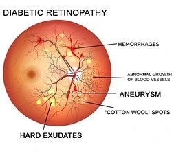
Diabetic retinopathy is a common eye complication caused by long-standing diabetes. High blood sugar levels damage the small blood vessels of the retina, leading to leakage, swelling, or abnormal vessel growth that can impair vision.
In its early stage, known as background diabetic retinopathy, patients may experience mild blurred or dim vision—especially if the macula (the central part of the retina) is affected. This stage often develops after 10–15 years of diabetes.
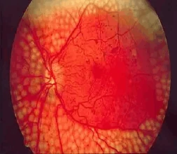
As the condition progresses, areas of the retina may stop receiving adequate blood supply, triggering the growth of fragile new blood vessels. This advanced stage, called proliferative diabetic retinopathy, can cause bleeding, scar formation, and even retinal detachment if left untreated.
Early consultation with a retina specialist in Pune is crucial for timely diagnosis, close monitoring, and effective treatment to preserve vision.
A detailed dilated retinal examination helps detect macular edema and proliferative changes. Advanced investigations such as fluorescein angiography and optical coherence tomography (OCT) are used to identify subtle retinal damage.
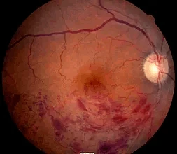
Retinal vein occlusion occurs when one of the tiny retinal veins becomes blocked by a blood clot. Risk factors, apart from advanced age and genetic factors, are smoking, obesity, high blood pressure, diabetes and high cholesterol levels. The occlusion of the vein prevents the drainage of blood which results in hemorrhages and a swelling of the surrounding retina. In the long run the retina is irreversibly damaged.
During this phase, guidance from Dr. Ramesh Murthy at Axis Eye Clinic—a highly experienced best retina surgeon in pune—is invaluable in ensuring precise diagnosis, modern treatment, and long-term visual care.
Age-related macular degeneration (AMD or ARMD) is one of the most common causes of vision loss in those aged over 50 years. AMD is a condition that occurs when cells in the macula degenerate and die. Damage to the macula affects the central vision which is needed for reading, writing, driving, recognizing people’s faces and doing other fine tasks. The disease does not lead to complete blindness. Visual loss can occur within months, or over many years, depending on the type and severity of AMD.
In this type the cells in the RPE of the macula gradually become thin (they ‘atrophy’) and degenerate. This layer of cells is crucial for the function of the rods and cones which then also degenerate and die. Typically, dry AMD is a very gradual process and patients may not totally lose their reading vision.
Wet AMD, also called neovascular AMD, is more aggressive and can cause rapid vision loss. Abnormal blood vessels grow beneath the retina and leak fluid or blood, leading to scarring of the macula and severe central vision impairment.
AMD is diagnosed through a detailed retinal examination by an ophthalmologist. Additional investigations may include:
Fundus photography
Fluorescein angiography
Optical coherence tomography (OCT)
With advanced diagnostics, personalized treatment plans, and expert care by an experienced retina specialist in Pune, Axis Eye Clinic is a trusted destination for comprehensive retina services. The clinic is committed to preserving vision and improving quality of life through early detection and modern retinal treatments.
Most people with diabetic retinopathy do not have any symptoms or visual loss due to their retinopathy. Initial symptoms that may occur include blurred vision, seeing floaters and flashes, or even having a sudden loss of vision. Without treatment, diabetic retinopathy can gradually become worse and lead to visual loss or even blindness.
As many patients are not aware of the disease even when advanced damage is present, it is very important for diabetics to have a regular, complete eye and retinal examination with dilation of the pupil. Treatment for diabetic eye disease is better at preventing and controlling the diabetic retinopathy than at reversing it once it is well established.
Laser seals leaking blood vessels to reduce macular edema, helping to prevent further vision loss. It also slows or stops growth of abnormal blood vessels, decreasing the chance of bleeding in the eye.
Retinal vein occlusion is a common cause of vision loss. It is most common in people over the age of 60 and it seems to affect both sexes equally.
The exact reason why a blood clot may form in one of the retinal veins is not clear. However, there are some things that are thought to increase your risk of developing retinal vein occlusion. They include the following
1. High blood pressure
2. Diabetes
3. Smoking
4. Cardiac diseases (heart related diseases)
5. Raised intraocular pressure (glaucoma)
© 2026. Axis Eye Clinic | All Rights Reserved