Retina is the light sensitive layer of cells at the back of the eye. It converts light into electrical signals which are sent to the brain through the optic nerve and the brain interprets them to produce the images that we see. A healthy retina is necessary for good vision.
To examine the retina, the ophthalmologist will dilate your eyes by using dilating drops. A retina specialist then uses a special magnifying lenses to examine your retina. The dilatation will reverse after 4-5 hours.
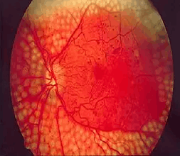
Diabetes can cause blood vessels to leak, causing fluid accumulation in the inner layers of the retina and thus affecting the vision. Abnormal blood vessels can also grow and possibly bleed. The damage and changes seen in the retina due to diabetes is called diabetic retinopathy.
A dilated retinal examination can reveal macular edema and proliferative diabetic retinopathy. Fluorescein angiogram and ocular coherence tomography (OCT) are useful in detecting subtle changes.
In mild cases, treatment for diabetic retinopathy is not necessary. Regular eye exams are critical for monitoring progression of the disease. Strict control of blood sugar and blood pressure levels can greatly reduce or prevent diabetic retinopathy. In more advanced cases, treatment is recommended to stop the damage due to diabetic retinopathy, prevent vision loss and restore vision.
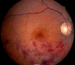
Retinal vein occlusion occurs when one of the tiny retinal veins becomes blocked by a blood clot. Risk factors, apart from advanced age and genetic factors, are smoking, obesity, high blood pressure, diabetes and high cholesterol levels. The occlusion of the vein prevents the drainage of blood which results in hemorrhages and a swelling of the surrounding retina. In the long run the retina is irreversibly damaged.
Treatment is aimed at preventing and treating any complications of the vein occlusion and control of risk factors. There is currently no treatment that can reverse the blocked vein.
Someone with retinal vein occlusion needs close follow-up so that any complications can be picked up early and treated where possible. Anti VEGF therapy in the form of injections needs to be given in case there are complications like neovascularization. Recurrence is real risk hence control of the underlying risk factors if of great importance.
Age-related macular degeneration (AMD or ARMD) is one of the most common causes of vision loss in those aged over 50 years. AMD is a condition that occurs when cells in the macula degenerate and die. Damage to the macula affects the central vision which is needed for reading, writing, driving, recognizing people’s faces and doing other fine tasks. The disease does not lead to complete blindness. Visual loss can occur within months, or over many years, depending on the type and severity of AMD.
In this type the cells in the RPE of the macula gradually become thin (they ‘atrophy’) and degenerate. This layer of cells is crucial for the function of the rods and cones which then also degenerate and die. Typically, dry AMD is a very gradual process and patients may not totally lose their reading vision.
Wet AMD may also be called neovascular or exudative AMD. It may cause severe visual loss within a short period – sometimes just months. Rarely if there is a bleed (hemorrhage) from a new blood vessel, this visual loss can occur suddenly, within hours or days. In wet AMD the retinal pigment cells degenerate and new tiny blood vessels grow from the tiny blood vessels in the choroid. This is called choroidal neovascularization. These vessels are fragile and tend to leak blood and fluid. This can damage the rods and cones, and cause scarring in the macula, causing severe vision loss.
Certain risk factors can lead to the development of AMD. These include:
ARMD is usually diagnosed after an eye specialist (an ophthalmologist) examines the retina at the back of the eye, using an ophthalmoscope. The retina at the back of your eye has a typical appearance in ARMD.
Special investigations may be performed to evaluate the extent of damage and this include:
1. Fundus photographs
2. Fluorescein angiography
3. Optical coherence tomography
Most people with diabetic retinopathy do not have any symptoms or visual loss due to their retinopathy. Initial symptoms that may occur include blurred vision, seeing floaters and flashes, or even having a sudden loss of vision. Without treatment, diabetic retinopathy can gradually become worse and lead to visual loss or even blindness.
As many patients are not aware of the disease even when advanced damage is present, it is very important for diabetics to have a regular, complete eye and retinal examination with dilation of the pupil. Treatment for diabetic eye disease is better at preventing and controlling the diabetic retinopathy than at reversing it once it is well established.
Laser seals leaking blood vessels to reduce macular edema, helping to prevent further vision loss. It also slows or stops growth of abnormal blood vessels, decreasing the chance of bleeding in the eye.
Retinal vein occlusion is a common cause of vision loss. It is most common in people over the age of 60 and it seems to affect both sexes equally.
The exact reason why a blood clot may form in one of the retinal veins is not clear. However, there are some things that are thought to increase your risk of developing retinal vein occlusion. They include the following
1. High blood pressure
2. Diabetes
3. Smoking
4. Cardiac diseases (heart related diseases)
5. Raised intraocular pressure (glaucoma)